Polyp Segmentation
U-Net with a Dice + BCE loss, tested at a second hospital; a screening threshold catches 97 % of polyp pixels.
Outlines colorectal polyps pixel by pixel in colonoscopy images with a U-Net (ResNet-18 encoder, ImageNet-pretrained), trained on a laptop CPU. Polyps are precursors of colorectal cancer; automatic segmentation helps endoscopists not miss them. Beyond the usual Dice score, the project measures what matters for a screening tool: test-time augmentation, a decision threshold tuned for sensitivity on validation data, and generalization to another hospital (CVC-ClinicDB).
No endoscopy images are shown: both datasets (Kvasir-SEG, CVC-ClinicDB) are licensed for research / education only and do not allow redistribution. All figures below are charts computed from them.
Results
All numbers come from one retrained model (docs/results.json): 20 epochs on CPU, best checkpoint at
epoch 17 (validation Dice 0.888). Kvasir-SEG test = 100 held-out images never used for training,
checkpoint selection or threshold choice. CVC-ClinicDB = 612 frames from another hospital, never seen.
Same center (Kvasir-SEG test) and the effect of test-time augmentation
| Kvasir-SEG test, threshold 0.5 | no TTA | flip TTA (4 views) |
|---|---|---|
| Mean Dice | 0.886 | 0.892 |
| Mean IoU | 0.823 | 0.829 |
| Median Dice | 0.942 | 0.946 |
| Pixel sensitivity / precision | 0.909 / 0.867 | 0.922 / 0.863 |
| Missed polyps (< 50 % of the polyp found) | 5 % | 3 % |
| Inference (CPU, 256×256) | 92 ms / image | 363 ms / image |
TTA adds +0.005 Dice on average, but image by image it helps 20 test images and hurts 19 (by more than 0.01 Dice): a small, noisy gain for 4× the compute. On CVC-ClinicDB it adds nothing (0.825 → 0.825).
Screening threshold (chosen on validation, reported on test)
A missed polyp is worse than a false alarm, so the threshold is chosen on the validation split to maximize pixel-level F2 (sensitivity counts 4× more than precision). Validation picked 0.05.
| Flip TTA | threshold 0.5 (default) | threshold 0.05 (F2 on validation) |
|---|---|---|
| Kvasir test: pixel sensitivity | 0.922 | 0.972 |
| Kvasir test: pixel precision | 0.863 | 0.713 |
| Kvasir test: mean Dice | 0.892 | 0.832 |
| Kvasir test: missed polyps | 3 % | 2 % |
| CVC-ClinicDB: pixel sensitivity | 0.804 | 0.903 |
| CVC-ClinicDB: pixel precision | 0.898 | 0.696 |
| CVC-ClinicDB: mean Dice | 0.825 | 0.779 |
| CVC-ClinicDB: missed polyps | 10.6 % | 4.4 % |
The low threshold finds more of every polyp and, more importantly, more than halves the polyps missed on the unseen hospital (10.6 % → 4.4 % of frames), at the cost of 2.7× (Kvasir) to 4.3× (CVC) more false-alarm area and about 5 Dice points. Dice is the wrong objective for screening; this table shows the trade-off explicitly.
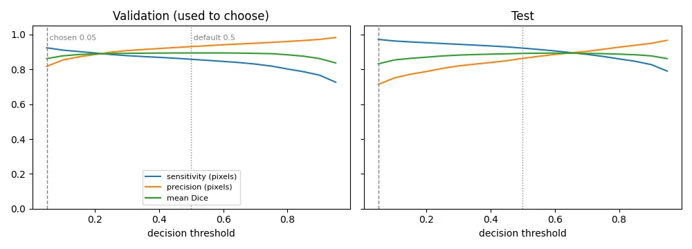
Sensitivity, precision and mean Dice against the decision threshold. Left: validation, where the threshold is chosen. Right: test, only scored afterwards; the curves have the same shape.
Another hospital (CVC-ClinicDB)
| Flip TTA, threshold 0.5 | Kvasir-SEG test (same center) | CVC-ClinicDB (other center) |
|---|---|---|
| Mean Dice | 0.892 | 0.825 |
| Mean IoU | 0.829 | 0.748 |
| Median Dice | 0.946 | 0.914 |
| Images with Dice > 0.8 | 84 % | 77 % |
| Images with Dice < 0.5 (failures) | 4 % | 9 % |
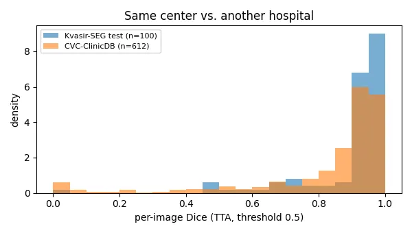
Moving to a different hospital, endoscope and resolution (384×288 frames instead of Kvasir’s mostly larger ones) costs about 7 Dice points on average. The median barely moves: most frames are still segmented well, but the failure tail doubles (4 % → 9 % of images below Dice 0.5), and sensitivity drops more than precision (0.92 → 0.80 vs 0.86 → 0.90): the model under-segments unfamiliar polyps rather than hallucinating new ones.
Reproducing the original baseline
The original weights were lost with a USB key, so the model was retrained from scratch with the same script, split and hyper-parameters:
| Row | Original run (README v1) | Retrained (this run) |
|---|---|---|
| Test mean Dice / IoU (no TTA) | 0.897 / 0.833 | 0.886 / 0.823 |
| Test median Dice | 0.937 | 0.942 |
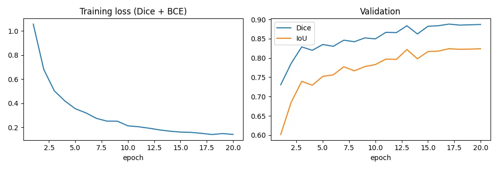
| Best validation Dice (epoch) | 0.882 (14) | 0.888 (17) |
| Time per epoch | ~3.5 min | 3.3–4.6 min (CPU shared with other jobs) |
The retrained model is 0.011 Dice lower on test but 0.006 higher on validation. The original run used unseeded augmentations (since fixed) and was interrupted in its last epoch, so it cannot be replayed exactly. With 100 test images and a per-image Dice standard deviation of roughly 0.15–0.2, the standard error of the mean is roughly 0.015–0.02, so the gap is within run-to-run noise. The 92 ms vs 69 ms inference difference comes from the laptop running other agents’ work at the same time.
For reference, published U-Net baselines on Kvasir-SEG report Dice of roughly 0.8–0.9 depending on split and resolution; specialized architectures reach the low 0.9s. Different papers use different splits, so treat this as context, not a head-to-head comparison.
How it works
- Data: Kvasir-SEG, 1,000 images with expert masks, resized to 256×256, split 800 / 100 / 100 (train / validation / test) with a fixed seed. CVC-ClinicDB (612 frames from another hospital) is used only for evaluation.
- Model: U-Net. The encoder (ResNet-18, ImageNet weights) extracts features at 5 scales, and
the decoder upsamples back to full resolution with skip connections carrying fine detail
(polyp boundaries) from the encoder. Built with
segmentation_models_pytorch. - Loss: Dice + binary cross-entropy. Polyps often cover < 20 % of the image. Pure BCE rewards predicting “background” everywhere, while the Dice term directly optimizes overlap with the polyp. BCE keeps gradients stable early on.
- Augmentation: flips and 90° rotations (applied identically to image and mask; colonoscopy has no canonical orientation), plus brightness/contrast jitter for endoscope lighting.
- Training: AdamW, cosine learning-rate decay; the checkpoint with the best validation Dice is kept.
- Test-time augmentation (TTA): the image is also predicted horizontally flipped, vertically flipped and both; each prediction is flipped back and the 4 probability maps are averaged.
- Screening threshold: thresholds 0.05–0.95 are swept on the validation split (with TTA) and the one maximizing pixel-level F2 (sensitivity weighted 4× over precision) is kept. The test split and CVC-ClinicDB are only scored with it afterwards.
Data
| Dataset | Role | Source | Licence |
|---|---|---|---|
| Kvasir-SEG (Jha et al., MMM 2020) | train / val / test | official zip, https://datasets.simula.no/downloads/kvasir-seg.zip (44 MB) | research and education only; cite the paper |
| CVC-ClinicDB (Bernal et al., CMIG 2015) | second-center test only | public Hugging Face file mirror berkaytrhn/cvc-clinicdb (612 PNG frames + masks, ~20 MB) | research and education only, commercial use forbidden; images © Hospital Clinic Barcelona, masks © Computer Vision Center Barcelona; cite the paper |
The official CVC-ClinicDB page no longer shows a working download link (the original was a .rar on
Google Drive), so the files come from a mirror that keeps the same 612 frames and masks and points
back to the official terms. Both datasets need no login. python download_data.py fetches both into
../../cv-data/kvasir/ (outside the repo) and deletes the archive after extraction.
Run it
# from the repo root, with the shared environment active (torch CPU + segmentation-models-pytorch)
cd 11-medical-segmentation
python download_data.py           # Kvasir-SEG + CVC-ClinicDB -> ../../cv-data/kvasir/
python train.py --epochs 20       # ~70 min on an idle CPU -> ../../cv-data/kvasir/outputs/best.pt
python evaluate.py                # TTA, tuned threshold, CVC-ClinicDB -> docs/results.json + charts
What I learned / what didn’t work
- TTA is not free accuracy here. The “+1 Dice point for free” folklore gave +0.5 point on Kvasir (with as many images hurt as helped) and nothing on CVC-ClinicDB, for 4× the inference time. The training augmentations already include the same flips, so the model is close to flip-invariant.
- The threshold matters more than TTA for screening. Going from 0.5 to 0.05 raised sensitivity on the unseen hospital from 0.80 to 0.90 and cut missed polyps from 10.6 % to 4.4 % of frames.
- The chosen threshold sits at the edge of the sweep (0.05). F2 on validation is nearly flat between 0.05 and 0.15 (0.900 vs 0.896), and an even lower threshold might score slightly higher. A finer grid near 0 or a target-sensitivity rule (“lowest threshold keeping precision above X”) would be a more stable choice; I kept the pre-registered grid rather than tuning it after seeing the result.
- Cross-center drop is mostly a failure tail. Median Dice falls only 0.03, mean Dice 0.07: a minority of CVC frames fail badly. CVC-ClinicDB’s 612 frames come from only ~29 video sequences, so near-duplicate frames make the failures look more frequent (and the sample bigger) than they are.
- Worst Kvasir cases are data artifacts. Some images carry a black inset box (an endoscope position overlay) that hides part of the frame and, in the worst case, almost the whole polyp. A real system would detect and mask out such overlays.
- False positives on folds: bulging mucosal folds look like flat polyps; lowering the threshold for sensitivity makes these false alarms more frequent (false-alarm area 2 % → 6 % of the image on test).
- Validation Dice plateaus after ~13 epochs (0.88–0.89) while training loss keeps falling.
- Not done: stronger encoder / higher resolution. The laptop’s CPU is shared by several projects
under a one-job-at-a-time lock; this single 77-min training waited many hours for its slot. A 352 px
ResNet-18 run would take about 2× longer per epoch (~2.5 h for 20 epochs), so it is left for a Kaggle
GPU session:
python train.py --size 352 --tag r18_352thenpython evaluate.py --size 352 --tag r18_352(both scripts already support it, plus--encoder efficientnet-b3).
Tech stack
Python · PyTorch · segmentation_models_pytorch (U-Net, ResNet-18) · NumPy · Matplotlib